Know more about the signs, symptoms and management of Tennis Elbow
Pain around the elbow is quite a common phenomenon. The pain is experienced particularly post vigorous activities or even during rest. Elbow joint is a joint, which connects the arm with forearm. It is composed of various bone ligaments, mascular, vascular and neural structures. Each structure around the elbow joint contributes to one specific pathology even the presentation is different.
The elbow has four sides- Superior (upper), Inferior (Lower side), Medial (Inner) and Lateral (Outer) surfaces.
The medial (inner) surface bears the nerve (Ulna), which gives sensation to the middle of the forearm, hand and fingers. The whole inner side of forearam muscles get attached to the radial aspect of elbow. Similarly, the outer muscle of forearm gets attached to the lateral aspect of elbow. (Humerus). The elbow joint is formed as Ulna humeral joint, Radio-ulna and Radio –humeral joint. The most common problem is Tennis Elbow in comparison to the Golfer’s elbow or any injury to the nerves.

What is Tennis Elbow?
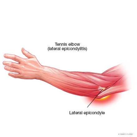
Tennis elbow is a condition, which affects the lateral aspect of the elbow joint. As the name suggests, it was commonly seen among tennis players due to repeated use of wooden racquet in earlier times.
Clinical Feature
Pain is localised to the lateral aspect of the elbow predominantly on the lateral epicondyle of humerus. Patient complains of difficulty in lifting weights even a glass of water or to perform and turn movement of the forearm such as rinsing of towels or opening the lock. Pain radiates towards the shoulder. The pain persists for a few days to months. A proper diagnosis and active intervention can ease it faster.
Sign
Tenderness present on the bony prominence of lateral epicondyle of humerus or lateral aspect of elbow. Pain also arises on pressuring the muscle bulk near the elbow. There is occasional tenderness on the triceps body and scapula. Patients also complain of pain while applying resistance or dorsiflexion of the wrist.
X-Ray
Predominantly, it looks normal. There will be barely the presence of any Osteophyte or Calcification. Most of the elbow joint helps to detect any localised Oedema, intimal tear of the tendurus muscles or any bony spur.
Treatment
- Analysis and anti-inflammation play a pivotal role. Most importantly patient needs rest of the affected limb for minimum 2 weeks.
- Release of the forearam, arm and scapula muscles is highly essential.
- Proper sleep therapy and stretching exercise of the forearam is required.
- Steroids ease the pain and improve movement temporarily.
- Role of PRP (Platelet Rich Plasma): It is still debatable. It’s a painful process.
- Surgical release is rarely required.

