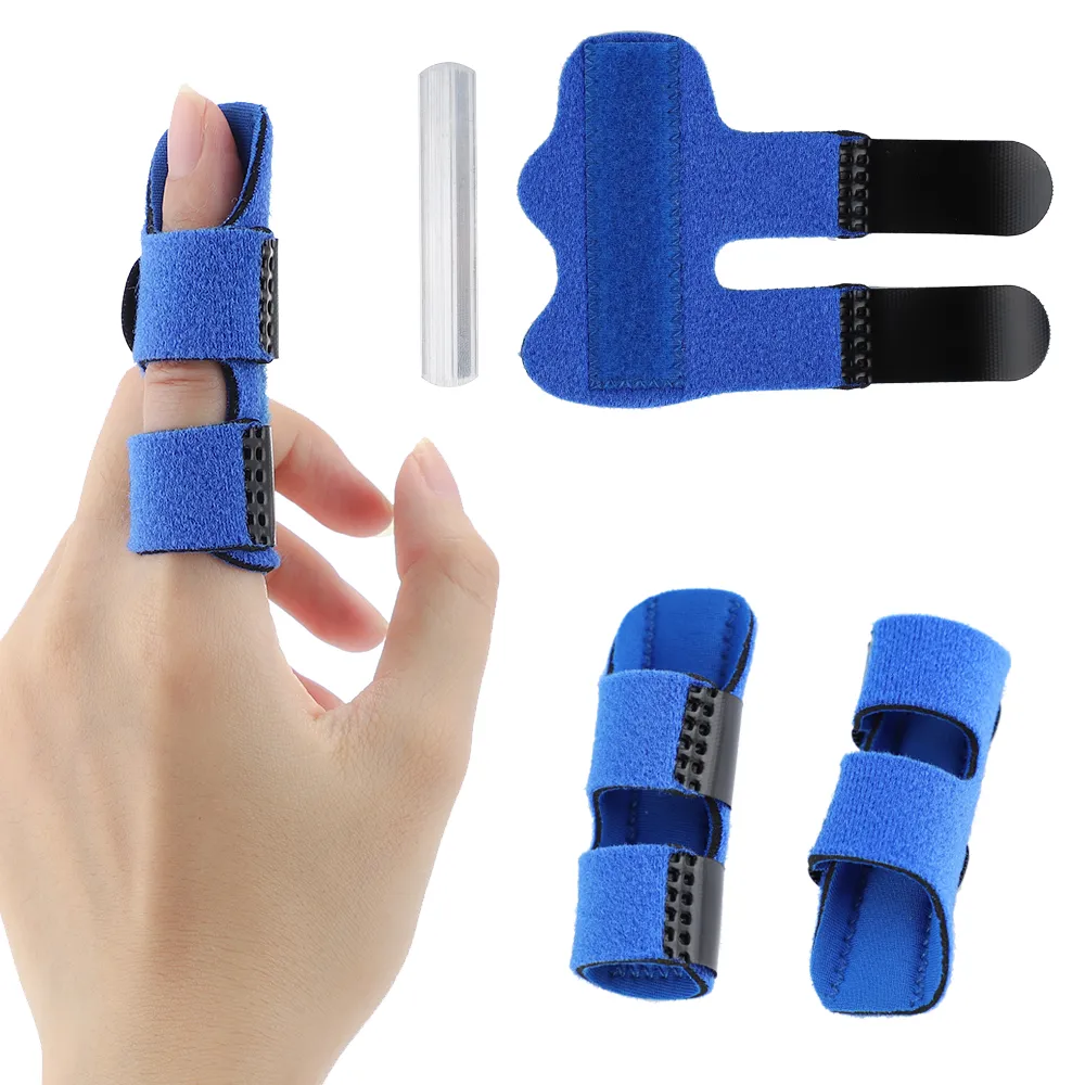
When the tendon gets torn or detached from the finger bone, the patient is unable to straighten the finger. And, that’s what is called the drop finger or mallet finger injury.
Have you ever heard of baseball finger injury? It is named so because it occurs commonly during the sport of baseball. However, it is also known as mallet finger injury. It occurs when the tip of a finger or the thumb is forcefully flexed. In this type of injury, the tendon that straightens the fingertip joint gets affected. Sometimes, it is also called the drop finger injury. Tendons attach bones to muscles. They provide stability and motion. So, when the tendon gets torn or detached from the finger bone, the patient is unable to straighten the finger. The finger—which looks bruised or swollen—droops at the tip. Hence, the name drop finger! The injury often affects a finger on the hand most frequently used. It is a common condition, especially in athletes. But it can also occur when performing household activities. If someone strikes the tip of a finger on an immovable object, then such an injury is experienced. You can get this injury even while performing simple tasks like arranging the bed.

Learn the anatomy
The fingers are made up of three bones called phalanges. The phalanges are separated by two joints. The distal interphalangeal (DIP) joints are located near the fingertips. The proximal interphalangeal (PIP) joints are located in the middle of the fingers. Extensor tendons are attached to the phalanges .These tendons allow the fingers to extend or straighten. In case of mallet finger injury, the extensor tendon that is attached to the distal phalanx gets torn due to the application of force. In some cases, the tendon may remain intact, but a small piece of bone can be pulled away from where it attaches to the phalanx. This is known as avulsion fracture. A patient can also face mallet finger if the extensor tendon is cut.
Symptoms of mallet finger
After experiencing the initial pain due to the injury, the patient may also witness the following symptoms:
- Swelling.
- Bruising.
- Redness.
- An inability to straighten fingertips.
- Tenderness.
- A detached fingernail.
- Redness under the fingernail bed.
It may also be noted that mallet finger is unrelated to arthritis. When the injury involves only the extensor tendon, subsequent arthritis doesn’t develop, usually. However, arthritis can occur if the tendon pulls off a piece of bone from the joint surface and remains displaced. It is very important to seek immediate attention, in case someone develops this injury. It must be remembered that if there is blood beneath the nail or if the nail gets detached, there is a possibility of developing infection.
CLASSIFICATION
According to Patel and Gerberman, mallet fingers can be classified as those presenting within 4 weeks of injury and the chronic ones (those presenting after 4 weeks of injury.) The most acceptable classification system for bony mallet finger is known as the Wehbe and Schneider classification system. In this system, the injury is divided into three types and each type is subdivided into three subtypes. The classification has been made depending on the degree of articular involvement.

Wehbe and Schneider classification
Types
- No DIP joint subluxation
- DIP joint subluxation
- Epiphyseal and physeal injuries
Subtypes
- Less than 1/3 of articular surface involvement
- 1/3 to 2/3 of articular surface involvement
- More than 2/3 of articular surface involvement
Diagnosis
The healthcare provider may require an X-ray of the affected part to detect the injury. On rare occasions, supplemental imaging studies such as an ultrasound or MRI may be needed.
Treatment
Immediately after the injury, first-aid is important. The victim should wrap an ice pack in a towel and put it on the affected finger. Hold the finger above your heart. Over-the-counter pain medications can be consumed, if needed.
Long-term treatment involves putting the fingertip into a splint. It should be kept there for a minimum of six weeks while the tendon heals. If there is a piece of bone pulled off, the healthcare provider may opt for another X-ray after a week or two of splinting. It is done to check on the appropriate position of the bone fragment and the healing process. The person may be needed to wear the splint day and night for at least six weeks. Avoid strenuous activities and sports to prevent recurrent injury. If the injury is more complex, a surgeon may need to surgically insert a small pin into the finger to hold the joint straight as it heals.
[/ihc-hide-content]


